
By Rayna Randers and Katy Cano; Farnsley Middle School (Louisville, KY)
When Katy (co-author) was born, her parents noticed a little red dot below her left eye. They thought it would eventually go away. However, the red dot didn’t go away and it kept getting bigger, up to half of her face causing her not to be able to see out of her left eye.
Katy’s parents, Aura Aguilar and Clementino Cano, states “We were worried so we took her to the hospital. We wanted to know what was happening to her face.”
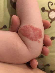
Katy’s doctor diagnosed her with hemangioma. According to the Mayo Clinic, “Hemangioma is a birthmark that commonly appears as a rubbery, bright red nodule of extra blood vessel in the skin.” Certain Hemangioma look darker than others and in some rare cases it to grows into the lower layers of the skin or in the muscle. This is called a deep lesion. In Katy’s case her lesion was only skin deep. “Hemangioma is extremely rare, 200,000 people are diagnosed each year,” stated the Mayo Clinic.
In an email to Scijourner, Leah Baker, who is part of The Disease and Vascular Malformation Team at the Cincinnati Children’s Hospital Medical Center, wrote that “In most cases a diagnosis is made by correlating medical history with a physical examination findings. In certain cases, radiological test such as Magnetic Resonance Imaging (MRI) or computed tomography may be used when trying to make a diagnosis or determining a lesion. It’s an imaging procedure that uses special x-ray equipment to create detailed pictures, or scans, of areas inside the body.”
The Mayo Clinic stated that some symptoms of hemangioma may be present at birth but more often appears during the first several months of life. It starts out as a flat red mark anywhere on the body most often on the face, scalp, chest, or back. Usually, a child has only one mark like Katy. Some children may have more than one mark particularly if they’re part of a multiple birth.
Katy’s doctors at the University of Florida Physicians requested to do laser surgery when Katy was about 3 months old. They told Katy’s parents that in some cases of hemangioma can disappeared or go away, but in Katy’s case her spot was half way across her face. Therefore, her birthmark would not have gone away.
The surgeries they recommended were done by using an extremely hot, focused beam of light that helps remove tissue and controls any bleeding from the surgery wound. Before she had the surgeries, her left eye was shut and Katy couldn’t see. Half of her face looked like a huge red balloon that was about to pop. In reality these surgeries helped her open her eye.
“We were worried that Katy would lose her eyesight,” states Katy’s parents.
According to the Hemangioma and Vascular Malformation Team, some common treatment options include drug therapy, which is a medical therapy used when the lesion is rapidly forming. Some medications include Timolol, which is a medication applied directly to the hemangioma, and corticosteroids, which can be injected into the skin or taken orally.
Complications of a hemangioma can lead to destruction of the skin and also develop a sore. This can also lead to pain and bleeding. Depending where the hemangioma is located, you may have trouble with vision, breathing, or hearing, but this usually rare.
Hemangioma is noncancerous and it really doesn’t increase your risk of getting cancer. And this tumor is usually starts smaller, but it can grow much larger.
In Katy’s case, laser surgery was needed due to her age. But there was some risk that it might result with a scar and changes in the area. Katy now has a scar underneath her left eye from all the surgeries.
“I now can see better and I am thankful to all doctors who helped me recover from Hemangioma,” says Katy. “Now I only have a scar on my left cheek and no longer have to go through surgeries”.

This work is licensed under a Creative Commons Attribution-NonCommercial-NoDerivs 3.0 Unported License.


